
Health & Wellness
InHealth: Mohs Micrographic Surgery
By Joseph F. Greco, M.D. | March 2023
What is Mohs Micrographic Surgery?
Earning the name from its pioneer, Dr. Frederic Mohs, the Mohs micrographic surgery procedure involves surgically removing skin cancer that we can clinically see in precise, thin layers and then microscopically examining it to make sure there’s no residual tumor remaining. That process repeats itself until the skin cancer is totally removed. One hundred percent of the peripheral and deep margins are examined leaving the smallest possible defect, and subsequent smallest possible scar. Considered to be the gold standard for treating skin cancer, Mohs micrographic surgery is hailed for its ability to spare as much healthy tissue as possible and may yield success rates up to 99%, the highest potential for most skin cancers.
Who is a candidate for Mohs Surgery?
Candidates for Mohs surgery include individuals with biopsy proven skin cancers, such as basal cell carcinoma and squamous cell carcinoma, in cosmetically sensitive areas such as the scalp, face, neck, hands and feet; skin cancers that are large or have histologically aggressive features seen under the microscope among other indications.
The importance of fellowship training
Why does this matter? A fellowship-trained Mohs surgeon must undergo a highly competitive application, review, and selection process to be chosen for training. Training includes a 1-to-2-year period of extensive and intensive hands-on direction and education from highly qualified surgeons who are themselves skilled practitioners of the Mohs procedure. During this period the fellow will be exposed to rare tumor pathology, difficult tumor locations, and highly complex wound reconstruction to build a unique and comprehensive surgical skillset.
The Procedure
A fellowship-trained Mohs surgeon will function in all phases of the Mohs surgery procedure and serve as the oncologic surgeon, dermatopathologist, and reconstructive surgeon on the same day.
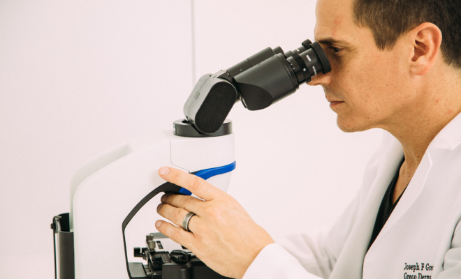
The area to be treated will be cleansed and local anesthesia will be administered. The visible tumor will be precisely removed along with a thin layer of surrounding tissue. This tissue will be taken immediately to the Mohs lab, processed, and placed onto slides so that it can be assessed by the surgeon under a microscope.
If evidence of cancer is seen, it will be precisely noted and matched to the exact location on the skin and another layer of tissue will be removed in the same manner. Layer after layer of tissue may be examined until no more cancer is detected within the tissue. This process ensures that only cancerous tissue is removed, which preserves as much healthy tissue as possible.
During the period while the tissue is being processed, the patient waits comfortably and privately in the exam room, with the ability to watch TV, listen to music, read, or quietly relax.
What is done once the cancer is removed?
Once the skin cancer is completely removed, your Mohs surgeon will immediately discuss the next steps to address the surgical wound with you. Small, superficial wounds may be allowed to heal on their own while some may be closed in a simple side-to-side manner. More challenging defects may require a skin graft or skin flap to preserve functional capabilities and maximize the aesthetic outcome. Reconstruction and closure are most often performed immediately after the skin cancer is clear.”
Joseph F. Greco, M.D., medical director of Greco Dermatology (grecodermatology.com), is a double-board certified dermatologist and fellowship trained Mohs surgeon. During his 15-year period at UCLA, Dr. Greco performed over 20,000 surgical procedures and participated in the training of 14 Mohs micrographic surgery fellows.

You must be logged in to post a comment Login